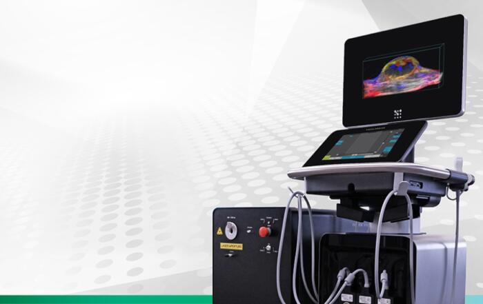
In this exciting webinar, Drs. Kristiina Aasa and Izzie Newsome will share details about the launch of an all-new imaging mode on the Vevo F2 system, Shear Wave Elastography Mode!
Elastography is an established method for visualizing and assessing liver fibrosis and tissue stiffness in the clinic. This powerful tool is now available at high frequency, specifically optimized for small animal imaging. Following this presentation, there will be a live Q&A Session where audience questions will be addressed.
Key Topics Include:
- Describe Shear Wave Elastography
- Discuss the value of Elastography in the clinic, and preclinically
- Showcase Elastography with live imaging of a mouse liver
- Share images of normal vs fibrotic livers
Presenters
Product Manager
FUJIFILM VisualSonics, Inc.
Kristiina Aasa completed her M.Sc. in Anatomy and Ph.D. in Biomedical and Molecular Sciences, both at Queen's University. She joined FUJIFILM VisualSonics 10 years ago as an Applications Scientist and is now the Product Manager.
Scientific Applications Specialist
Fujifilm VisualSonics, Inc.
Dr. Isabel (Izzie) Newsome is a Scientific Applications Specialist for Fujifilm VisualSonics. In 2021, she completed her Ph.D. in biomedical engineering at the University of North Carolina at Chapel Hill in the lab of Dr. Paul Dayton, where her dissertation focused on novel contrast-enhanced ultrasound methods and nonlinear microbubble dynamics in the context of tumor-associated angiogenesis.
Webinar Host
FUJIFILM VisualSonics Inc.
FUJIFILM VisualSonics designs and manufactures ultra high frequency in vivo imaging systems, for both research and clinical use. our ultrasound platform provides images at resolutions that far exceed any other system available on the market. Beyond ultrasound, we have also developed a unique photoacoustic technology to expand on the capabilities of our imaging solutions.

Additional Content From FUJIFILM VisualSonics Inc.
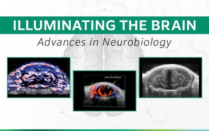
Illuminating the Brain: Advances in Neurobiology
Watch leading experts in neurobiology imaging in a virtual symposium exploring cutting-edge multi-modal approaches using ultra-high frequency ultrasound and photoacoustics.
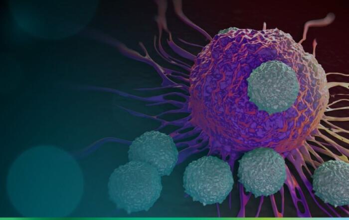
The Sound of Science: Mapping the Tumor Microenvironment for Advanced Therapeutic Strategies
Unlock the full potential of photoacoustic imaging (PAI) for evaluation of the tumor microenvironment at VisualSonics' Advanced Photoacoustic Imaging Workshop.
Vevo Ask The Experts: Oncology Imaging
Experts at FUJIFILM VisualSonics explore how the Vevo F2 can advance oncology research through high-resolution ultrasound and photoacoustic imaging.
Related Content
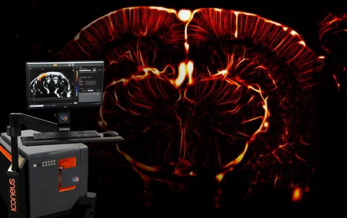
Functional Ultrasound Demonstration and Live Q&A: Iconeus One, a Pioneer in Real-Time Whole-Brain Imaging
Join us for an online session to discover the capabilities of functional ultrasound (fUS) with the Iconeus One system.
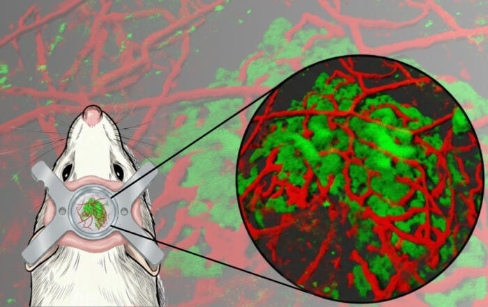
Opening a New Window: Stable Long-Term Imaging of Dura Mater–Transplanted Pancreatic Islets in Awake Mice
Join Drs. Tröster and Köhler as they present a novel dura mater transplantation platform for long-term, anesthesia-free imaging of pancreatic islets in awake mice.
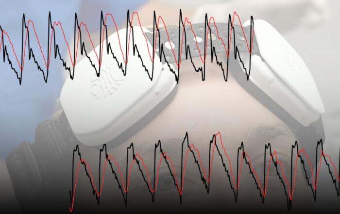
Clinical Applications of Optical Cerebral Blood Flow Imaging
Join Dr. Chris Favilla as he discusses non-invasive measurement of cerebral blood flow and potential applications.