Join Dr. Chris Albanese and Dr. Olga Rodriguez for this webinar to discuss Ultrasound Photoacoustic Imaging (US-PAI) and how it can directly detect key liver disease markers.
Chronic diseases of the liver, including liver steatosis and steatohepatitis, are associated with extensive fibrosis, which is typically performed by needle biopsy.
Current widely used imaging approaches for liver diagnosis exist but do not provide sufficient diagnostic accuracy for defining the various stages of fibrosis or steatosis.
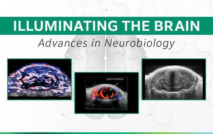
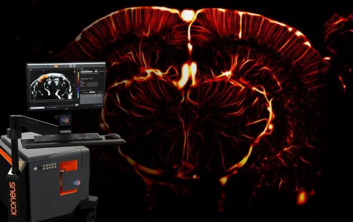
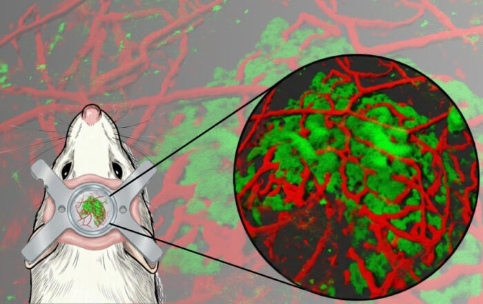
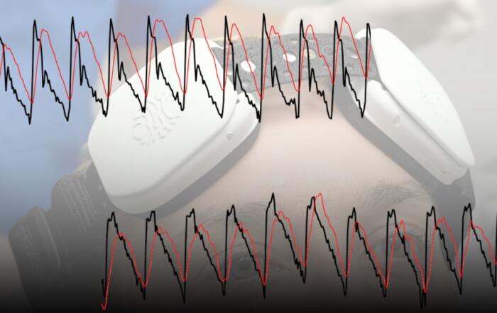
Ultrasound-based Photoacoustic Imaging (US-PAI) has recently emerged as a non-invasive, non-ionizing modality, capable of capturing structural details and oxygen saturation changes during disease progression. However, its potential for detecting surrogate MASLD markers, such as collagen and lipids, had not been investigated in detail.
This study used innovative spectral photoacoustic imaging for the direct detection and quantification of key biomarkers of liver disease, such as fibrosis, collagen, lipids, oxygenated and deoxygenated hemoglobin, establishing that US-PAI, validated with MRI, effectively identified increases in liver adiposity and fibrosis in a preclinical model of liver disease.
Key Topics Include:
- Liver diseases like steatosis and fibrosis are usually diagnosed with needle biopsy, which is invasive. Current imaging methods can’t reliably show the different stages of disease
- Ultrasound Photoacoustic Imaging (US-PAI) is a new, non-invasive approach that captures tissue structure and oxygen changes
- How US-PAI can directly detect key liver disease markers like collagen, fat, and fibrosis
- Results, confirmed with MRI, suggest US-PAI could become a powerful tool for tracking liver disease without biopsy
Presenters
Professor of Oncology and Radiology
Director, Preclinical Imaging Research Laboratory (PIRL) and Executive Director, Center for Translational Imaging (CTI)
Georgetown University Medical Center

Associate Professor of Oncology
Co-Director – Preclinical Imaging Research Laboratory (PIRL)
Georgetown University Medical Center
Production Partner
FUJIFILM VisualSonics Inc.