In this webinar, Dr. Mickael Tanter presents his work on functional ultrasound neuroimaging, including an overview and translation to clinical applications.
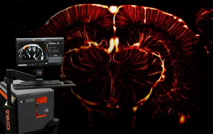
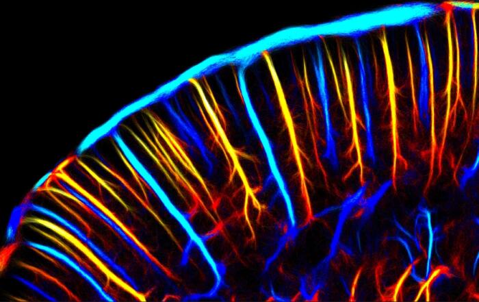
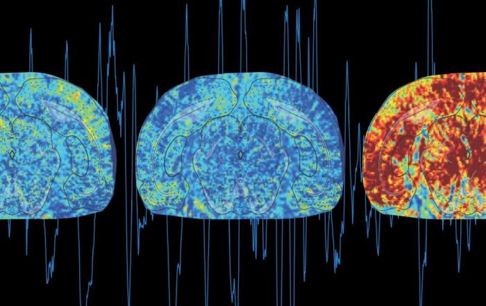
Functional ultrasound (fUS) was first introduced by Dr. Mickael Tanter in a landmark paper in 2011. Since this first publication, and as a result of technical advancements and scientific ingenuity, fUS methods have progressed rapidly and have now been used in a wide range of applications, including neuroimaging. In this webinar, Dr. Tanter presents an overview of his research on fUS, including its key features and applications, using examples from a number of neuroscience studies. He also discusses functional ultrasound localization microscopy (fULM), and how preclinical applications of fUS can be translated to the clinic.
Key Topics Include:
- Overview of the key differentiating features of functional ultrasound in the field of neuroimaging, illustrated by neuroscience studies
- How to translate from preclinical fUS imaging to clinical applications
- Functional ultrasound localization microscopy (fULM): the how and why of functional ultrasound at a microscopic scale
- Categories: Microscopy, Neuroscience, Preclinical Imaging, Vascular Biology
- Tags: fMRI, functional ultrasound, functional ultrasound (fUS), imaging, in vivo imaging, Neuroimaging, neurovascular, neurovascular coupling, preclinical imaging, vascular function, vascular imaging
Presenters

Mickael Tanter, PhD
Professor/Research Director
Physics for Medicine Paris
Inserm
Production Partner
Iconeus

Additional Content From Iconeus
Functional Ultrasound Demonstration and Live Q&A: Iconeus One, a Pioneer in Real-Time Whole-Brain Imaging
Iconeus One: Pioneering Real-Time Whole-Brain Imaging with Functional Ultrasound – 14 Years of Innovation and Adoption
Multimodal Neuroimaging Using Functional Ultrasound (fUS)
Having issues registering with our form? Please Register Here