Q&A Report: Reprogramming the Brain Tumor Microenvironment to Enhance Anti-Tumor Immunity
What do you think makes the macrophage infiltrate into tumors better than CD8+ T cells?
There must be chemokines which influence macrophage trafficking but we also see that macrophages express high levels of some matrix metalloproteases, which probably help break down the ECM to allow for easier access to the surrounding tissue.
Is there a shift in anti-inflammatory macrophage proportions in tumor recurrence? Thank you.
Early data suggests that recurrent GBM tissue shows more immunosuppressive macrophage cells compared with de novo GBM.
How heterogenous is the macrophage population in glioblastoma? Do you have any data on macrophage subsets during disease progression?
We have only started to look at macrophage heterogeneity but don’t have clear data yet. We see the 3 subsets I mentioned based on the antibodies used but the cells will exist across a continuum from anti-inflammatory to pro-inflammatory.
Thanks for the nice presentation. How long does it take for a pro-inflammatory, anti-tumor macrophage to become an anti-inflammatory, pro-tumor macrophage? For example, if I would like to test a new cancer treatment, would 24h of treatment be enough to see a shift in the macrophage activation status?
We can only judge timing to convert an M1-like state to M2 via in vitro experiments & RNA-seq & proteomics. We know that immusuppressive gene mRNA in macrophages are seen after 8h & proteins after 24h. Based on cell signaling changes seen in macrophages, macrophages can activate the immusuppressive signals in about 20 minutes of stimulation by GBM cell-conditioned medium.
What are some ways to overcome TME and make immunotherapy more effective?
Dampening the M2-like pro-tumor macrophages is one way. Activating T-cells directly by targeting checkpoint inhibitors has been one concept showing promise in some cancers but has not worked for brain cancer. Overcoming T-cell exhaustion to maintain longer anti-tumor response has been another aim of some research & this has largely been focussed on targeting specific transcription factors in T-cells, such as TOX & FOXO.
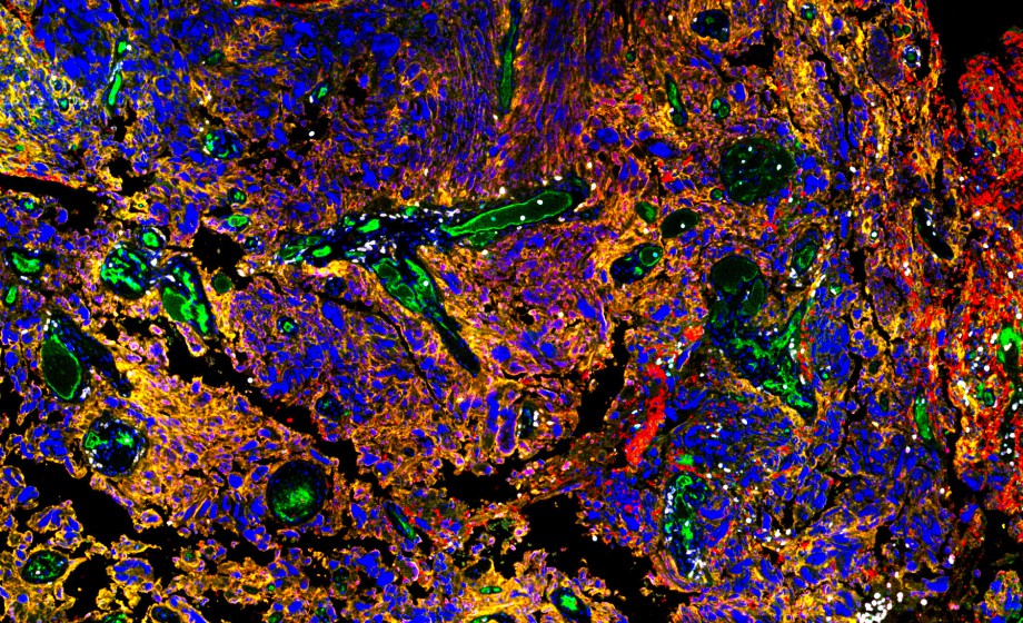
Great talk! Have you performed additional experiments to show that the collagen is why the T cells are "trapped"? Have you seen any relevant immunosuppressive interaction in this sense? Thank you.
Great question. We believe that the pathological levels of perivascular ECM deposition is one reason for lack of efficient immune cell trafficking but we also believed there are specific cytokines & chemokines in these niches which influence trafficking. We don’t yet know which of these are important but there are some candidates we have identified.
What is the significance of CD47 in the MAC/Microglia panel? Does it have/hold/play an essential role in GBM response to immunotherapy?
CD47 is the so-called “don’t eat me” (anti-phagocytosis) signal, expressed on the cancer (in fact all/most) cell membrane, so it is a potential anti-cancer target, where blocking CD47 could allow more efficient phagocytosis of cancer cells by macrophages.
Thanks for your talk, is there a difference in immune infiltration in angiogenic areas versus co-opted (hijacked) brain vessels?
Good question. It looks like most of the “trapped” immune cells are in more mature regions of the tumor rather than neo-angiogenic regions, which have thinner perivascular regions.
Were any of these biomarkers examined in relation to any treatments given either in mouse models or in humans?
Some of the multiplex antibody panels have PD-1 & PDL1 antibodies, which are targets.
What is the immune response in Alzheimer's disease?
I have not investigated the immunology of AD. A good review discussing AD immunopathology is: https://doi.org/10.1016%2Fj.immuni.2022.10.016
Did you check TGF-β signaling in GBM and its impact on myeloid cells?
No, we haven’t looked at TGF-β signaling but we have detected TGF-β in the GBM TME.
Do you have information on cytokine expression in the TME as well? Have you looked at FOXP3 expression too?
We see very few FoxP3+ Tregs in brain tumors. We have managed to see lots of IL10 in the brain tumor TME, using multiplex IHC & we suspect that the neoplastic cells & macrophages are the main cells producing IL10.
Do you expect to see a difference in cAMPi sensitivity between newly diagnosed and recurrent GBM?
Interesting question. We have not looked at cAMP signaling directly but CREB, a major downstream target of cAMP signaling appears to be more highly activated in recurrent GBM.
Have you noticed increased difficulty with visualization due to nonspecific binding to endogenous antigen as you increase in the number of biomarkers detected in these assay? How have you overcome these challenges as you increase to large biomarker numbers?
We have had some difficulty with background on some tissues but this seems to be dependent on the tissue sample. For 7-color mIHC we have optimized the antibody concentration & the antigen retrieval buffer pH (either pH ~7 or ~9) using standard chromogenic IHC first. The order of antibody staining can make a difference. For the higher-plex (>15 antibody) platform, we have not had major problems because the antibodies are pre-optimized by the manufacturer.
How do you ensure the medium is accurate to what is found in a patient? Are there any microenvironment changes or components that are lost/gained that could be overlooked by capturing it in just one time point?
Good question. In our studies so far, we have examined many patient tissues (>150) so we’re confident of the data & it’s statistically validated when comparing disease/tumor stage/age/other parameters. One should consider what the question is & determine how many samples one needs to be confident that observations/stats are robust. For longitudinal studies, where a patient has multiple tumor biopsies taken over time, again one would need to have enough patients to be confident of data. Having said that, for biological phenomena, e.g. cell-ECM interaction, fewer samples would be needed but you would want to examine larger tissue sections to account for intra-tissue heterogeneity.
Hi! Great talk! Is it worth it to do complex spatial experiments in mouse tumors? The models I have been working on (B16 melanoma, MC38 colon cancer) grow quickly and do not show much tumor niche heterogeneity. Human tumors in contrast do show a lot of intratumoral heterogeneity, maybe due to months/years of tumor evolution/immunoediting. Opinion/ideas to overcome this issue?
I agree that most/all mouse models for brain cancer are probably too rapid, so don’t exhibit similar early chronic pathology, as is the case in human brain tumors. We use multiplex IHC for mouse models to examine cell composition to compare to human tumors, as well as examine changes in immune cell trafficking in response to pharmacological intervention, to understand mechanisms underpinning this.
Great results, thank you. Have you done similar research on meningioma and have similar findings?
We have not looked at meningioma but a recent paper which has is: https://doi.org/10.1038/s41588-024-01747-1
What type of collagen do you see in the perivascular niche (Type I, Type IV)?
Mainly collagen 1 & 4 – we have not performed a focussed ECM mIHC protein analysis yet – some of this data is in our paper: https://doi.org/10.1007/s13402-022-00763-9
Thank you for the talk. Could you comment on the importance of NK cells on the immune landscape of GBM? Can this cell type, together with T cell activation increase immune response in GBM or other brain cancers scenario?
NK cells must have a role in tumor immunity but we have not looked at this, as we’ve had problems with NK cell-specific antibodies.
What is astrocytes' role in tumor suppression?
We have not studied the role of healthy astrocytes in brain tumors. One of the best papers investigating this is: https://doi.org/10.1038/s41467-019-10493-6